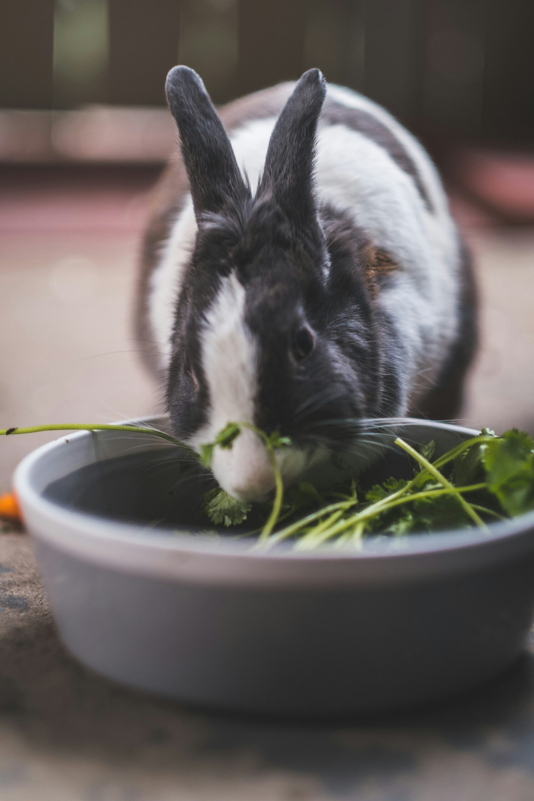
Rabbit’s bodies are well equipped to live on low quality foods, such as grasses and weeds. To gain nutrition from these items, they have a specialized gastrointestinal tract that allows them to break down this fibrous roughage and turn it into usable energy. Much of the work is done by a large section of the intestine called the cecum, where the fibrous plant material is fermented and broken down by a complex microbiome of bacteria and yeast. But the work begins in the mouth.
Rabbits have hypselodont dentition, meaning teeth that grow constantly throughout life and have long crowns. If rabbit’s teeth weren’t constantly growing, they would be worn down by all the plant material consumed over time. Because the teeth are constantly emerging (like our fingernails), the situation in a rabbit’s mouth is dynamic and can change dramatically within their lifetime. When the teeth are wearing down normally, they don’t appear to be changing at all; but if the teeth start to have problems, disease can set in quite quickly as the teeth are growing several millimeters every week.
In a normal rabbit, there are six incisors that create a chisel to help chew through food. Once food enters the mouth, the twenty-two “cheek teeth” grind on the hay in a circular motion, breaking down the fiber into digestible sizes.

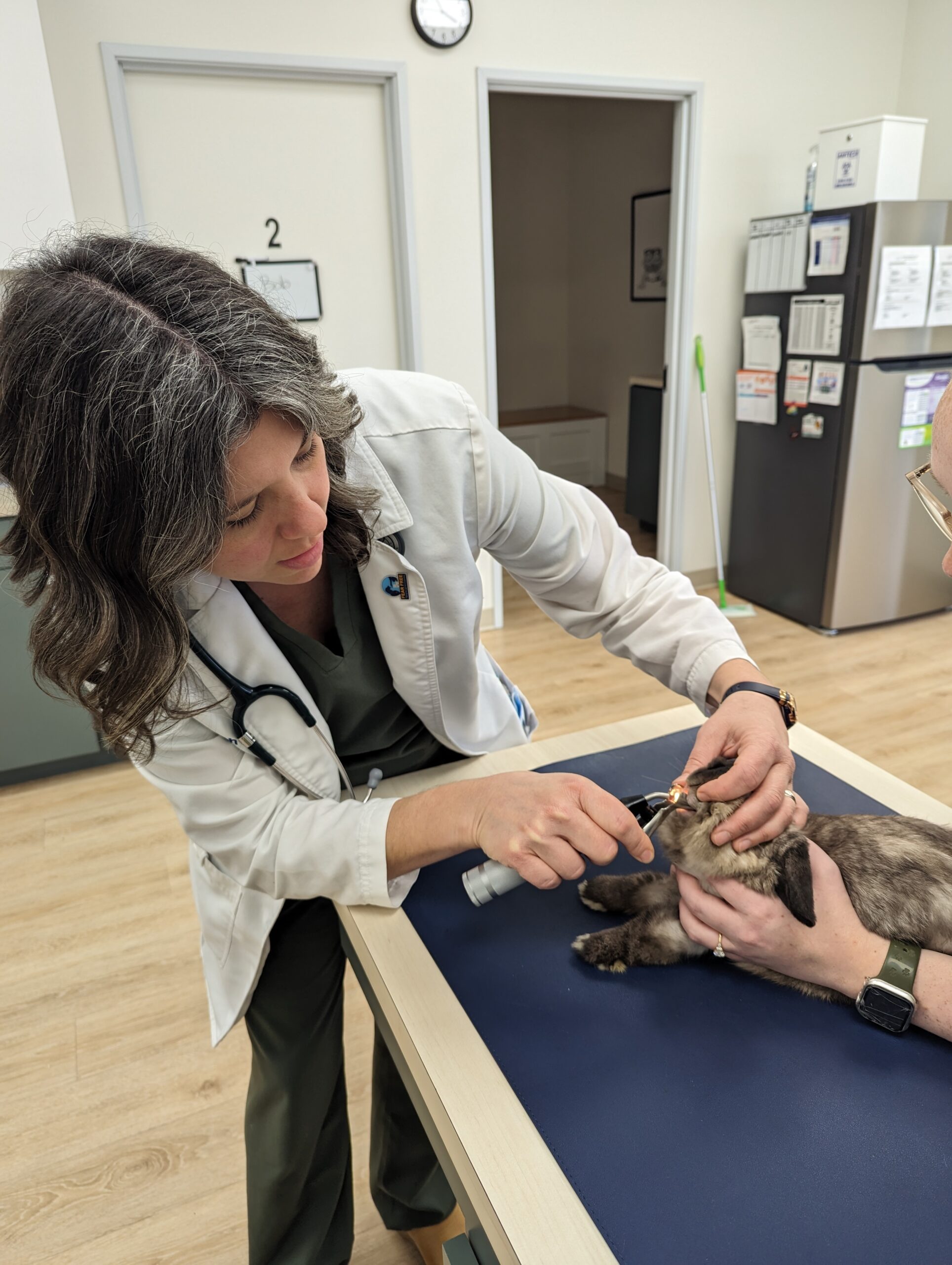
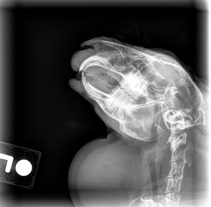
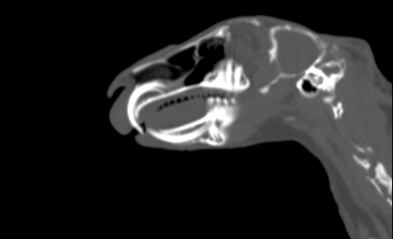
There are a number of issues frequently seen in rabbits:
Overgrown incisors: If the incisors are not wearing on each other appropriately, they can become overgrown and result in ulceration of the mucus membranes. The upper incisors can also curl into the mouth and cause serious trauma to the roof of the mouth. Overgrown incisors can result from an underbite at birth, or secondary to disease of the cheek teeth.

Cheek teeth spurs/points: Normal cheek teeth naturally have a slight angle to them, but diseased teeth will grow sharp spurs. These spurs are typically found near the tongue on the lower teeth, or along the cheeks on the upper teeth. Sharp spurs can cause pain with eating, as well as ulceration of the tongue and gum tissue.

Dental abscesses: Diseased teeth are also prone to infection. Often this infection will be present within the jaw bone, undetected by owners. When the infection eventually wears through the bone, an abscess (a large mass on the face or jaw filled with pus) will rapidly emerge. It’s important to remember that the abscess is not the only problem, but rather a symptom of dental disease. Antibiotics will NOT resolve the abscess. Instead, the swelling must be treated surgically and the underlying dental disease diagnosed and treated, often with extraction of the offending teeth.
Depending on the type of dental disease, there are several treatments that may be necessary:
Filing of cheek teeth spurs: If there are sharp points on the teeth, they can be trimmed or filed slightly to provide immediate relief to a patient. This is typically done with sedation and may need to be followed with additional dental work.
Molar float: Performed under anesthesia, a molar float involves using a high speed bur to reduce the length of the teeth in the mouth. The goal is to get the teeth back to normal position and allow the mouth to close normally once again.
Dental extractions: Diseased and infected teeth may need to be removed. Specialized instruments are used to carefully extract the teeth. Since most of the tooth is buried below the surface, sometimes an extraoral approach (an incision and removal of bone from the outside) is necessary to remove diseased teeth.
Abscess treatment: Abscesses associated with dental disease must be opened and disease tissue removed. The abscess site is typically kept open after surgery to allow for ongoing topical wound care.
Preventing dental disease in rabbits is far more rewarding, easier on the patient, and significantly less expensive. The most important way to keep your rabbit’s teeth healthy is to provide an appropriate diet. Adult rabbits should have unlimited access to high quality Timothy or Orchard grass hay. Plain, homogenous pellets such as Oxbow Adult Rabbit Food, Purina Fiber3, or Mazuri Rabbit Diet should be given daily, about ¼ cup for most rabbits (giant breeds can have ½ cup). Additionally, rabbits can have a handful of leafy greens every day. Carrots, fruits, and treats should almost never be fed. If you do give a rabbit fruit, a serving size is about one blueberry and should not be given daily.
